
CD1a Antibody (DA016)
CD1a Antibody (DA016)
Recombinant monoclonal rabbit antibody
Description
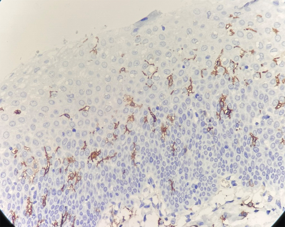
CD1a is a non-polymorphic, major histocompatibility complex, class I-related cell surface glycoprotein (45 to 55 kDa) and is expressed in association with β-microglobulin.1 CD1a is predominantly expressed in dendritic cells and thymic cortical cells, and is positive in 70% of thymocytes, and is often not expressed in early thymic cells or mature cells. In normal tissues, anti-CD1a reacts with cortical thymocytes, Langerhans cells, interdigitating cells, and rare antigen-presenting cells of the lymph node.2 CD1a positivity has also been seen in Langerhans cell histiocytosis (histiocytosis X)3, and a subset of pre-T lymphoblastic lymphoma/leukemia (cortical T LBL/L).4,5 CD1a is used for the classification of thymoma, and cortical thymoma; CD1a can be used for the differentiation of thymic tumors from primary lung tumors.
References
2. Angel CE, et al. Distinctive localization of antigen-presenting cells in human lymph nodes. Blood. 2009; 113:1257-67.
3. Emile JF, et al. Langerhans' cell histiocytosis. Definitive diagnosis with the use of monoclonal antibody O10 on routinely paraffin-embedded samples. Am J Surg Pathol. 1995; 19:636-41.
4. Stefano, AP et al. Acute leukemia immunophenotyping in bone-marrow routine sections. Br J Haematol. 1999; 105:394-401.
5. Han X, et al. Precursor T-cell acute lymphoblastic leukemia/lymphoblastic lymphoma and acute biphenotypic leukemias. Am J Clin Pathol. 2007; 127:528-44.
Specifications
Order information
| RMB1A055 | 0.1ml, 0.5ml, 1ml | Concentrate |
| RMB1A055 | 3ml, 6ml, 10ml | Ready to use |
