
EMA Antibody (DA055)
EMA Antibody (DA055)
Recombinant monoclonal rabbit antibody
Description
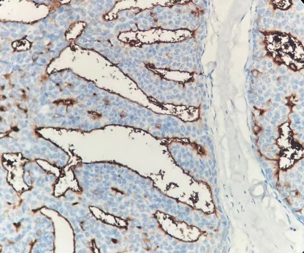
EMA is a transmembrane glycoprotein with a molecular weight of 400 kDa that is widely distributed in epithelial cells and tumors of epithelial origin. Anti-EMA antibody is a useful marker for staining many carcinomas. It stains normal and neoplastic cells from various tissues, including mammary epithelium, sweat glands and colorectal carcinoma. Hepatocellular carcinoma, adrenal carcinoma and embryonal carcinomas are consistently EMA negative, so keratin positivity with negative EMA favors one of these tumors. EMA is frequently positive in meningioma, which can be useful when distinguishing it from other intracranial neoplasms such as schwannomas.1-8
References
1. Pinkus GS, et al. Epithelial membrane antigen--a diagnostic discriminant in surgical pathology: immunohistochemical profile in epithelial, mesenchymal, and hematopoietic neoplasms using paraffin sections and monoclonal antibodies. Hum Pathol. 1985; 16:929-40.
2. Pinkus GS, et al. Are keratin proteins a better tumor marker than epithelial membrane antigen? A comparative immunohistochemical study of various paraffin-embedded neoplasms using monoclonal and polyclonal antibodies. Am J Clin Pathol. 1986; 77:269-77.
3. Dearnaly DP, et al. Increased detection of mammary carcinoma cells in marrow smears using antisera to epithelial membrane antigen. Br J Cancer. 1981; 44:85-90.
4. Redding WH, et al. Detection of micrometastases in patients with primary breast cancer. Lancet 1983; 1271-4.
5. Attanoos RL, et al. The use of immunohistochemistry in distinguishing reactive from neoplastic mesothelium. A novel use for desmin and comparative evaluation with epithelial membrane antigen, p53, platelet-derived growth factor-receptor, P-glycoprotein and Bcl-2. Histopathology. 2003; 43:231-8.
6. Beer TW, et al. Ber EP4 and epithelial membrane antigen aid distinction of basal cell, squamous cell and basosquamous carcinomas of the skin. Histopathology. 2000; 37:218-23.
7. Lee JS, et al. Immunohistochemical panel for distinguishing between carcinoma and reactive mesothelial cells in serious effusions. Acta Cytol. 1996; 40:631-6.
8. Fraga M, et al. Bone marrow involvement in anaplastic large cell lymphoma. Immunohistochemical detection of minimal disease and its prognostic significance. Am J Clin Pathol. 1995; 103:82-9.
Specifications
Application:
IHC (FFPE)
Host:
Rabbit
Dilution range:
Immunogen:
Synthetic peptide of human EMA
Cellular location:
Cytoplasm/Cell membrane
Control:
Normal Breast
Order information
| MMB1A067 | 3ml, 6ml, 10ml | Ready to use |
